Protanopia

People with protanopia have no working red-sensitive (L) cones. Reds appear much darker, and red, orange, yellow and green are seen as variations of a similar hue. Deep reds can look almost black.

Protanopia is the strongest form of protan color blindness, also called red-blindness. The milder form is protanomaly.

Red-green color vision deficiency · Last updated 2026-11-10

What does protanopia look like?
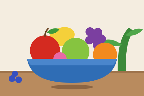
Illustration of fruit in a blue bowl with typical color vision: red apple, green apple, yellow lemon, orange, purple grapes, pink peach and blueberries.Typical color vision
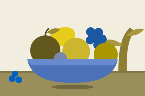
The same fruit bowl simulated with protanopia. The red apple turns dark olive brown, and red, orange, yellow and green collapse into similar olive and yellow tones. Purple looks blue and pink looks grayish blue.Simulated protanopia
The red apple turns dark olive brown, and red, orange, yellow and green collapse into similar olive and yellow tones. Purple looks blue and pink looks grayish blue.
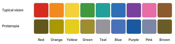
Nine color swatches (red, orange, yellow, green, teal, blue, purple, pink and brown) with typical vision and simulated with protanopia.
Common colors with typical vision (top) and simulated protanopia (bottom). Images are simulations based on the Machado, Oliveira & Fernandes (2009) model; real experience varies from person to person and with your screen.
  • Reds look dark, sometimes close to black.
  • Red, orange, yellow and green look similar.
  • Blue and yellow remain distinct.

Try our color blind simulator to see your own photos the way someone with a protan deficiency might.

How common is protanopia?

About 1% of men; very rare in women.

Everyday situations that can be tricky

  • Red brake lights and stop signs rely on shape and position more than color.
  • Red text or red error messages may be hard to notice.
  • Some occupations require normal color vision.

Causes

Inherited on the X chromosome. Lifelong and stable.

Protanopia vs protanomaly

Both are protan types: they involve the red-sensitive (L) cones. In protanopia those cones are missing or not working, so reds look dark and many reds, oranges and greens look the same. In protanomaly the cones are present but shifted, so the same confusions are milder and severity varies from person to person.

Read more about protanomaly.

Glasses, apps and other aids

Color-enhancing glasses are unlikely to help with protanopia because an entire cone type is missing. What the studies found, type by type: do color blind glasses work?

Phone and computer accessibility settings (color filters), color-identifier apps, and asking for labels or patterns on color-coded information can all help. There is currently no approved cure for inherited color blindness.

How is protanopia diagnosed?

Eye care professionals use plate tests (such as Ishihara or HRR plates), arrangement tests and, for a precise diagnosis, an anomaloscope. Online tests like ours can flag a likely deficiency but can’t replace a clinical test.

Take the red-green test

Protanopia FAQ

What is protan color blindness?

Protan color blindness is the group of red-green color vision deficiencies that involve the red-sensitive (L) cones: protanopia (missing or not working) and protanomaly (shifted). It is less common than deutan color blindness.

What does protanopia look like?

The images above simulate it: reds look much darker, and red, orange, yellow and green look like variations of a similar hue. Blue and yellow remain distinct.

Why do reds look dark with protanopia?

The red-sensitive cones contribute most to how bright long-wavelength (red) light looks. Without them, deep reds send a much weaker signal, so they can look dark brown or nearly black.

How common is protanopia?

About 1% of men; it is very rare in women.

Is there a protanopia filter?

Yes. Our color blind simulator applies a protanopia filter to your own photos in the browser, so you can see roughly how an image looks with protanopia.

Other types of color blindness

Protanopia is one of four types of red-green color blindness. See all types of color blindness compared side by side, or what colorblind people see with each type.

Sources

  1. National Eye Institute. Types of color vision deficiency. nei.nih.gov
  2. MedlinePlus Genetics, U.S. National Library of Medicine. Color vision deficiency. medlineplus.gov
  3. Birch J. Worldwide prevalence of red-green color deficiency. JOSA A. 2012;29(3):313–320. doi.org
  4. Nathans J, Piantanida TP, Eddy RL, Shows TB, Hogness DS. Molecular genetics of inherited variation in human color vision. Science. 1986;232(4747):203–210. pubmed.ncbi.nlm.nih.gov
  5. Simunovic MP. Colour vision deficiency. Eye. 2010;24(5):747–755. pubmed.ncbi.nlm.nih.gov
  6. Machado GM, Oliveira MM, Fernandes LAF. A physiologically-based model for simulation of color vision deficiency. IEEE TVCG. 2009;15(6):1291–1298. (Used for the simulated images.) doi.org

Note: this is a screening, not a diagnosis. Online tests can’t control your screen’s colors or brightness, so confirm any result with an eye care professional. Medical disclaimer · How the test works

More color vision tests